Can Hardware Removal be Avoided Using Bioresorbable Mg-Zn-Ca Screws After Medial Malleolar Fracture Fixation? Mid-Term Results of a First-In-Human Study
Abstract
Ankle is the most common site of hardware removal, mainly performed within 12 months of the primary surgery. The prominence of the metallic hardware is a frequent cause of pain after fracture fixation. Over the last decade, the development of bioresorbable materials based on magnesium (Mg) has increased. Bioresorbable metals aim to avoid a second surgery for hardware removal.
Methods
Twenty patients with isolated, bimalleolar, or trimalleolar ankle fractures were treated with bioresorbable screws made of Mg, 0.45wt% calcium (Ca) and 0.45wt% zinc (Zn) (ZX00). Patient-reported outcome measures (PROMs) including visual analogue scale (VAS) for pain, the presence of complications 6 and 12 months after surgery and the AOFAS scale after 12 months were reported. The functional outcomes were analysed through the range of motion (ROM) of the ankle joint with a standard goniometer. Degradation products and the bioresorbability of the screws were evaluated using plane radiographs.
Results
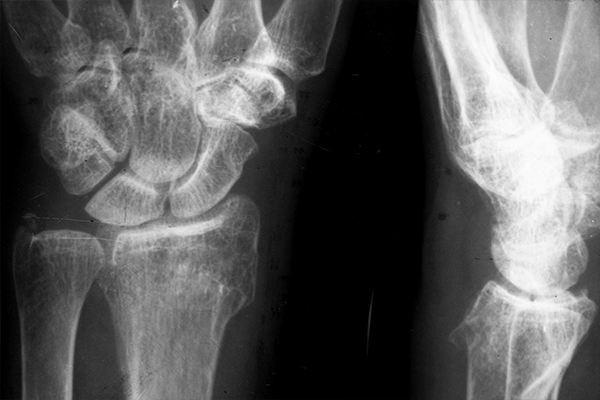
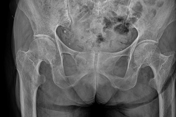
One patient was lost to follow-up. All patients were free of pain, no complications, shoe conflict or misalignement were reported after 12 months of follow-up. No Mg screws were surgically removed. An additional fixation of the distal fibula or the dorsal tibial fragment with conventional titanium implants (Ti) was performed in 17 patients. Within 12 months after primary refixation, 12 of these patients (71%) underwent a second surgery for Ti hardware removal. The mean AOFAS score was 89.8±7.1 and the difference between the treated and the non-treated site in the ROM of the talocrural joint was 2°±11° after 12 months. Radiolucent areas around the screws were attributed to degradation and did not affect clinical or functional outcomes. After one year, the Mg screw heads could not be detected in the plane radiographs of 17 patients which suggests that the majority of the screw head is degraded without introducing adverse reactions.
Conclusions
At 6 and 12 months, the bioresorbable Mg screws show excellent PROMs without complications or need for screw removal. The resorbability of the screw heads in most of the patients after one year could also provide an advantage over conventional bio-inert implants by avoiding related skin irritation due for instance to shoe conflict.
The combined effect of zinc and calcium on the biodegradation of ultrahigh-purity magnesium implants
Abstract
Magnesium (Mg)-based implants are promising candidates for orthopedic interventions, because of their biocompatibility, good mechanical features, and ability to degrade completely in the body, eliminating the need for an additional removal surgery. In the present study, we synthesized and investigated two Mg-based materials, ultrahigh-purity ZX00 (Mg-Zn-Ca; <0.5 wt% Zn and <0.5 wt% Ca, in wt%; Fe-content <1 ppm) and ultrahigh-purity Mg (XHP-Mg, >99.999 wt% Mg; Fe-content <1 ppm), in vitro and in vivo in juvenile healthy rats to clarify the effect of the alloying elements Zn and Ca on mechanical properties, microstructure, cytocompatibility and degradation rate. Potential differences in bone formation and bone in-growth were also assessed and compared with state-of-the-art non-degradable titanium (Ti)-implanted, sham-operated, and control (non-intervention) groups, using micro-computed tomography, histology and scanning electron microscopy. At 6 and 24 weeks after implantation, serum alkaline phosphatase (ALP), calcium (Ca), and Mg level were measured and bone marrow stromal cells (BMSCs) were isolated for real-time PCR analysis. Results show that ZX00 implants have smaller grain size and superior mechanical properties than XHP-Mg, and that both reveal good biocompatibility in cytocompatibilty tests. ZX00 homogenously degraded with an increased gas accumulation 12 and 24 weeks after implantation, whereas XHP-Mg exhibited higher gas accumulation already at 2 weeks. Serum ALP, Ca, and Mg levels were comparable among all groups and both Mg-based implants led to similar relative expression levels of Alp, Runx2, and Bmp-2 genes at weeks 6 and 24. Histologically, Mg-based implants are superior for new bone tissue formation and bone in-growth compared to Ti implants. Furthermore, by tracking the sequence of multicolor fluorochrome labels, we observed higher mineral apposition rate at week 2 in both Mg-based implants compared to the control groups. Our findings suggest that (i) ZX00 and XHP-Mg support bone formation and remodeling, (ii) both Mg-based implants are superior to Ti implants in terms of new bone tissue formation and osseointegration, and (iii) ZX00 is more favorable due to its lower degradation rate and moderate gas accumulation.
Degradation behavior and osseointegration of Mg-Zn-Ca screws in different bone regions of growing sheep—a pilot study
Abstract
Magnesium (Mg)-based implants are highly attractive for the orthopedic field and may replace titanium (Ti) as support for fracture healing. To determine the implant–bone interaction in different bony regions, we implanted Mg-based alloy ZX00 (Mg < 0.5 Zn < 0.5 Ca, in wt%) and Ti-screws into the distal epiphysis and distal metaphysis of sheep tibiae. The implant degradation and osseointegration were assessed in vivo and ex vivo after 4, 6 and 12 weeks, using a combination of clinical computed tomography, medium-resolution micro computed tomography (µCT) and high-resolution synchrotron radiation µCT (SRµCT). Implant volume loss, gas formation and bone growth were evaluated for both implantation sites and each bone region independently. Additionally, histological analysis of bone growth was performed on embedded hard-tissue samples. We demonstrate that in all cases, the degradation rate of ZX00-implants ranges between 0.23 and 0.75 mm/year. The highest degradation rates were found in the epiphysis. Bone-to-implant contact varied between the time points and bone types for both materials. Mostly, bone-volume-to-total-volume was higher around Ti-implants. However, we found an increased cortical thickness around the ZX00-screws when compared with the Ti-screws. Our results showed the suitability of ZX00-screws for implantation into the distal meta- and epiphysis.
Keywords: biodegradable implants, magnesium-based alloys, computed tomography, Mg–Zn–Ca, sheep, histology
In vitro and in vivo degradation behavior of Mg-0.45Zn-0.45Ca (ZX00) screws for orthopedic applications
Abstract
Magnesium (Mg) alloys have become a potential material for orthopedic implants due to their unnecessary implant removal, biocompatibility, and mechanical integrity until fracture healing. This study examined the in vitro and in vivo degradation of an Mg fixation screw composed of Mg-0.45Zn-0.45Ca (ZX00, in wt.%). With ZX00 human-sized implants, in vitro immersion tests up to 28 days under physiological conditions, along with electrochemical measurements were performed for the first time. In addition, ZX00 screws were implanted in the diaphysis of sheep for 6, 12, and 24 weeks to assess the degradation and biocompatibility of the screws in vivo. Using scanning electron microscopy (SEM) coupled with energy dispersive X-ray spectroscopy (EDX), microcomputed tomography (μCT), X-ray photoelectron spectroscopy (XPS), and histology, the surface and crosssectional morphologies of the corrosion layers formed, as well as the bone-corrosion-layer-implant interfaces, were analyzed. Our findings from in vivo testing demonstrated that ZX00 alloy promotes bone healing and the formation of new bone in direct contact with the corrosion products. In addition, the same elemental composition of corrosion products was observed for in vitro and in vivo experiments; however, their elemental distribution and thicknesses differ depending on the implant location. Our findings suggest that the corrosion resistance was microstructure-dependent. The head zone was the least corrosion-resistant, indicating that the production procedure could impact the corrosion performance of the implant. In spite of this, the formation of new bone and no adverse effects on the surrounding tissues demonstrated that the ZX00 is a suitable Mg-based alloy for temporary bone implants.
Keywords
Magnesium alloys, Biodegradable implants, Microstructure, Electron microscopy, Corrosion layers
Long-term in vivo degradation of Mg–Zn–Ca elastic stable intramedullary nails and their influence on the physis of juvenile sheep
Abstract
The use of bioresorbable magnesium (Mg)-based elastic stable intramedullary nails (ESIN) is highly promising for the treatment of pediatric long-bone fractures. Being fully resorbable, a removal surgery is not required, preventing repeated physical and psychological stress for the child. Further, the osteoconductive properties of the material support fracture healing. Nowadays, ESIN are exclusively implanted in a non-transphyseal manner to prevent growth discrepancies, although transphyseal implantation would often be required to guarantee optimized fracture stabilization. Here, we investigated the influence of trans-epiphyseally implanted Mg–Zinc (Zn)–Calcium (Ca) ESIN on the proximal tibial physis of juvenile sheep over a period of three years, until skeletal maturity was reached. We used the two alloying systems ZX10 (Mg-1Zn-0.3Ca, in wt%) and ZX00 (Mg-0.3Zn-0.4Ca, in wt%) for this study. To elaborate potential growth disturbances such as leg-length differences and axis deviations we used a combination of in vivo clinical computed tomography (cCT) and ex vivo micro CT (μCT), and also performed histology studies on the extracted bones to obtain information on the related tissue. Because there is a lack of long-term data regarding the degradation performance of magnesium-based implants, we used cCT and μCT data to evaluate the implant volume, gas volume and degradation rate of both alloying systems over a period of 148 weeks. We show that transepiphyseal implantation of Mg–Zn–Ca ESIN has no negative influence on the longitudinal bone growth in juvenile sheep, and that there is no axis deviation observed in all cases. We also illustrate that 95 % of the ESIN degraded over nearly three years, converging the time point of full resorption. We thus conclude that both, ZX10 and ZX00, constitute promising implant materials for the ESIN technique.
Keywords
Elastic stable intramedullary nails (ESIN), Physis, Growth plate, Biodegradable implants, Mg–Zn–Ca, Sheep study, Long-term degradation behavior, ZX00, ZX10, Pediatric fracture treatment
CORR Insights®: Mg-Zn-Ca Alloy (ZX00) Screws Are Resorbed at a Mean of 2.5 Years After Medial Malleolar Fracture Fixation: Follow-up of a First-in-humans Application and Insights From a Sheep Model
This CORR Insights® is a commentary on the article “Mg-Zn-Ca Alloy (ZX00) Screws Are Resorbed at a Mean of 2.5 Years After Medial Malleolar Fracture Fixation: Follow-up of a First-in-humans Application and Insights From a Sheep Model” by Labmayr and colleagues available at: DOI: 10.1097/CORR.0000000000002799.
Biodegradable ultrahigh-purity magnesium and its alloy ZX00 promote osteogenesis in the medullary cavity and glycogenolysis in the liver
Abstract
Magnesium (Mg)-based implants have become an attractive alternative to conventional permanent implants in the orthopedic field. While biocompatibility, degradation kinetics, and osseointegration of Mg-based implants have been mostly investigated, the impact of degradation products on bone remodeling and potential systemic effects remains unclear. The aim of this study was to evaluate the early and mid-term local and systemic tissue responses of degrading ultrahigh-purity ZX00 (Mg–Zn–Ca alloy) and ultrahigh-purity Mg (XHP-Mg) pins in a juvenile healthy rat model. The potential differences between implant types (degradable vs. permanent), implantation, and age-related changes were investigated using titanium (Ti), sham-operated, and control groups (non-intervention), respectively.
Degradation products of ZX00 and XHP-Mg pins promote osteogenesis in the medullary cavity by upregulating the expression levels of Bmp2 and Opg within 14 days post-surgery. The higher degradation rate of XHP-Mg resulted in the accumulation of degradation products starting from day 3 and upregulation of different genes, particularly Ccl2 and Cepbp. Besides good osseointegration and new bone tissue formation, we found a more parallel hydroxyapatite/collagen orientation along Mg-based pins in the perimeter region compared to Ti pins. In the liver, reduced glycogen levels in Mg-based pins indicated that degradation products promote glycogenolysis, while only the ZX00 group showed a higher serum glucagon level on day 14. Results suggest that degrading ZX00 and XHP-Mg pins stimulate osteogenesis mainly via Bmp2 and Opg and promote glycogenolysis in the liver, while the higher degradation rate of XHP-Mg pins resulted in upregulation of different genes and metabolites.
Statement of significance
Bioresorbable magnesium (Mg)-based implants are promising alternative candidates for orthopedic interventions. Until now, a few in vivo studies explored how Mg-based implants promote osteogenesis in the medullary cavity and modulate systemic tissue responses. Herein, results demonstrate i) the degradation rate of the Mg-based implants has a crucial effect on osteogenesis via regulating Bmp2 and Opg expression in the medullary cavity, ii) a parallel HAp/collagen matrix pattern in ZX00 and XHP-Mg groups compared to the Ti group, iii) both Mg pins promote glycogenolysis in the liver. Our findings highlight the dual role of Mg-based implants in bone remodeling and systemic metabolic modulation. Nevertheless, this is the first study to report the interaction between Mg-based implants and liver metabolism.
Mg-Zn-Ca Alloy (ZX00) Screws Are Resorbed at a Mean of 2.5 Years After Medial Malleolar Fracture Fixation: Follow-up of a First-in-humans Application and Insights From a Sheep Model
Background
In the ongoing development of bioresorbable implants, there has been a particular focus on magnesium (Mg)-based alloys. Several Mg alloys have shown promising properties, including a lean, bioresorbable magnesium-zinc-calcium (Mg-Zn-Ca) alloy designated as ZX00. To our knowledge, this is the first clinically tested Mg-based alloy free from rare-earth elements or other elements. Its use in medial malleolar fractures has allowed for bone healing without requiring surgical removal. It is thus of interest to assess the resorption behavior of this novel bioresorbable implant.
Questions/purposes
(1) What is the behavior of implanted Mg-alloy (ZX00) screws in terms of resorption (implant volume, implant surface, and gas volume) and bone response (histologic evaluation) in a sheep model after 13 months and 25 months? (2) What are the radiographic changes and clinical outcomes, including patient-reported outcome measures, at a mean of 2.5 years after Mg-alloy (ZX00) screw fixation in patients with medial malleolar fractures?
Methods
A sheep model was used to assess 18 Mg-alloy (ZX00) different-length screws (29 mm, 24 mm, and 16 mm) implanted in the tibiae and compared with six titanium-alloy screws. Micro-CT was performed at 13 and 25 months to quantify the implant volume, implant surface, and gas volume at the implant sites, as well as histology at both timepoints. Between July 2018 and October 2019, we treated 20 patients with ZX00 screws for medial malleolar fractures in a first-in-humans study. We considered isolated, bimalleolar, or trimalleolar fractures potentially eligible. Thus, 20 patients were eligible for follow-up. However, 5% (one patient) of patients were excluded from the analysis because of an unplanned surgery for a pre-existing osteochondral lesion of the talus performed 17 months after ZX00 implantation. Additionally, another 5% (one patient) of patients were lost before reaching the minimum study follow-up period. Our required minimum follow-up period was 18 months to ensure sufficient time to observe the outcomes of interest. At this timepoint, 10% (two patients) of patients were either missing or lost to follow-up. The follow-up time was a mean of 2.5 ± 0.6 years and a median of 2.4 years (range 18 to 43 months).
Results
In this sheep model, after 13 months, the 29-mm screws (initial volume: 198 ± 1 mm 3 ) degraded by 41% (116 ± 6 mm 3 , mean difference 82 [95% CI 71 to 92]; p < 0.001), and after 25 months by 65% (69 ± 7 mm 3 , mean difference 130 [95% CI 117 to 142]; p < 0.001). After 13 months, the 24-mm screws (initial volume: 174 ± 0.2 mm 3 ) degraded by 51% (86 ± 21 mm 3 , mean difference 88 [95% CI 52 to 123]; p = 0.004), and after 25 months by 72% (49 ± 25 mm 3 , mean difference 125 [95% CI 83 to 167]; p = 0.003). After 13 months, the 16-mm screws (initial volume: 112 ± 5 mm 3 ) degraded by 57% (49 ± 8 mm 3 , mean difference 63 [95% CI 50 to 76]; p < 0.001), and after 25 months by 61% (45 ± 10 mm 3 , mean difference 67 [95% CI 52 to 82]; p < 0.001). Histologic evaluation qualitatively showed ongoing resorption with new bone formation closely connected to the resorbing screw without an inflammatory reaction. In patients treated with Mg-alloy screws after a mean of 2.5 years, the implants were radiographically not visible in 17 of 18 patients and the bone had homogenous texture in 15 of 18 patients. No clinical or patient-reported complications were observed.
Conclusion
In this sheep model, Mg-alloy (ZX00) screws showed a resorption to one-third of the original volume after 25 months, without eliciting adverse immunologic reactions, supporting biocompatibility during this period. Mg-alloy (ZX00) implants were not detectable on radiographs after a mean of 2.5 years, suggesting full resorption, but further studies are needed to assess environmental changes regarding bone quality at the implantation site after implant resorption.
Clinical relevance
The study demonstrated successful healing of medial malleolar fractures using bioresorbable Mg-alloy screws without clinical complications or revision surgery, resulting in pain-free ankle function after 2.5 years. Future prospective studies with larger samples and extended follow-up periods are necessary to comprehensively assess the long-term effectiveness and safety of ZX00 screws, including an exploration of limitations when there is altered bone integrity, such as in those with osteoporosis. Additional use of advanced imaging techniques, such as high-resolution CT, can enhance evaluation accuracy.
A lean magnesium-zinc-calcium alloy ZX00 used for bone fracture stabilization in a large growing-animal model
Over the last decade, demand has increased for developing new, alternative materials in pediatric trauma care to overcome the disadvantages associated with conventional implant materials. Magnesium (Mg)-based alloys seem to adequately fulfill the vision of a homogeneously resorbable, biocompatible, load-bearing and functionally supportive implant. The aim of the present study is to introduce the high-strength, lean alloy Mg‒0.45Zn‒0.45Ca, in wt% (ZX00), and for the first time investigate the clinical applicability of screw osteosynthesis using this alloy that contains no rare-earth elements. The alloy was applied in a growing sheep model with osteotomized bone (simulating a fracture) and compared to a non-osteotomy control group regarding degradation behavior and fracture healing. The alloy exhibits an ultimate tensile strength of 285.7 ± 3.1 MPa, an elongation at fracture of 18.2 ± 2.1%, and a reduced in vitro degradation rate compared to alloys containing higher amounts of Zn. In vivo, no significant difference between the osteotomized bone and the control group was found regarding the change in screw volume over implantation time. Therefore, it can be concluded that the fracture healing process, including its effects on the surrounding area, has no significant influence on degradation behavior. There was also no negative influence from hydrogen-gas formation on fracture healing. Despite the proximal and distal screws showing chronologically different gas release, the osteotomy showed complete consolidation.
Statement of Significance
Conventional implants involve several disadvantages in pediatric trauma care. Magnesium-based alloys seem to overcome these issues as discussed in the recent literature. This study evaluates the clinical applicability of high-strength lean Mg‒0.45Zn‒0.45Ca (ZX00) screws in a growing-sheep model. Two groups, one including a simulated fracture and one group without fracture, underwent implantation of the alloy and were compared to each other. No significant difference regarding screw volume was observed between the groups. There was no negative influence of hydrogen-gas formation on fracture healing and a complete fracture consolidation was found after 12 weeks for all animals investigated.
Keywords
Degradation; Fracture fixation; Growing skeleton; Magnesium-based implants; Pediatric orthopedics